Conventional cell-biological TEM applications and correlative microscopy
Our group supports a broad range of “classical” cell-biological TEM methods such as ultra-thin sectioning of resin-embedded tissues, cells and isolated cellular components, immunogold-labelling methods, Tokuyasu-cryo-sectioning as well as metal-shadowing techniques In TEM, we collaborate with all departments and research groups of the institute having a wet lab but also with external collaboration partners. TEM sample preparation is technically difficult and time-consuming and, in most cases, requires careful optimization for each sample to achieve optimal image contrast depending on the nature of the tissue and cells, respectively. TEM projects are therefore performed as full-service projects and are considered as scientific collaboration rather than routine service, with many projects resulting in long-term collaborations.

One example for a long-term project is the analysis of the structure, assembly- and infection-mechanism of the bacterial phage SPP1 that infects the Gram-positive model bacterium Bacillus subtilis. Originally introduced by the funding director of the MPIMG, Thomas Trautner, SPP1 phages until today serve as a model system for eukaryotic dsDNA viruses and have been intensively studied in the institute over many years. After the retirement of Rudi Lurz (former head of the microscopy group) in 2012, we were continuing the close collaboration with Paulo Tavares (former member of the Trautner lab; CNRS, Gif-sur-Yvette) and have now applied Tokuyasu cryo-sectioning and immunogold electron microscopy to to study the spatiotemporal compartmentalization of viral components during virus replication in the bacterial cell. We could show that during infection, SPP1 procapsids localize at the periphery of the viral DNA compartment for genome packaging whereas DNA-filled capsids segregate into flanking warehouses that are temporally and spatially independent from the freshly replicated viral DNA (Labarde et al., 2021, PNAS).
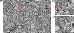
Conventional TEM on ultrathin-sectioned resin-embedded samples provides ultrastructural details at nanometer resolution. However, the field of view of a single TEM image is limited to some µm squared depending on the final magnification used, and finding certain biological events therefore often corresponds to the proverbial search for a needle in a haystack. We therefore implemented automated data acquisition based on the Leginon system (Suloway et al., 2005, J Struct Biol), which enables us to routinely image large regions of interest up to 500 x 500 µm2 at a pixel size of ~1-2 nm. The acquired micrographs (often more than thousand images per region) are then stitched using the TrakEM plugin in FIJI (Cardona et al., 2012, PLoS One) to one single montage (Figure 1) which can then be inspected for statistical analysis. In collaboration with Stephan Sigrist (FU-Berlin), we applied this approach to analyze statistically aging-related changes in the number of active zones as well as the density of synaptic vesicles per synaptic bouton in the calyx-region of drosophila melanogaster brains (Figure 1; Gupta et al., 2017, PLoS Biol).
This collaboration led to several follow-up projects, where automated TEM imaging was applied to study iPSC-derived neuronal stem cells (Lorenz et al., 2017, Cell Stem Cell), hepatocyte-like cells generated from human Induced pluripotent stem cells (Matz et al., 2017, Sci Rep) and cancer model systems (Brandt et al., 2019, Nat Commun; Bischoff et al., 2020, iScience). In ongoing projects, we currently use automated TEM to image early mouse embryo development at the blastocyst state (Bulut-Karslioglu lab), mouse brain development (Kraushar lab), cancer model systems (Dept. Meissner, Hnisz lab) as well as fibroblast cells derived from patients showing severe progeroid features (FG Mundlos).
A new focus of our service group is the implementation of correlative light and electron microscopy techniques. Recently, we used correlative approaches to study transmission of equine herpes viruses between peripheral blood mononuclear cells from the lung to endothelial cells (Kamel et al., 2020, iScience) and SPP1 phage assembly in bacteria (Labarde et al., 2021, PNAS). As test system, we also established a HEK cell spheroid model system, which can be used for high-throughput and 3D light microscopy as well as TEM and SEM imaging. Moreover, we aim at establishing robust image analysis workflows to analyze ultrastructural details such as organelles, vesicles, fat droplets or cell-cell contacts, amongst others (van der Weijden et al., 2022, bioRxix). Within a new project with the Kraushar lab, we are currently establishing pre-embedding immuno-staining protocols for quantification of ribosome populations during brain development.
